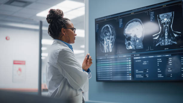
Correctly referred to as imaging, these studies aid in diagnosis and are usually performed by the Radiology department. Some other departments also perform imaging studies and this is a trend which will likely increase as new imaging methods are developed.
The Radiology department performs X-rays, Ultrasounds, CT Scans, PET Scans, MRI, Nuclear Medicine Scans and Fluoroscopy studies in addition to some others, depending on what needs there are for additional information from imaging.

New technologies have made it possible for images of the human body to be acquired which can assist doctors in their quest to arrive at accurate diagnoses. Images may be static, in which case they are like photographs and capture an event that is occurring at a particular moment in time or they may be dynamic/functional and capture a process that occurs over a longer time period. In either case they provide useful information that would not have been available in even the recent past, and when added to other information increase the likelihood of an accurate diagnosis.
Whenever images are obtained they are interpreted by a radiologist within a short time period and an official report is then generated (electronically or in hard copy) so that the findings can be shared with the doctor attending to the patient. The radiologist may also be asked to point out the finding(s) on the actual images as they may be subtle and not easily identified by an untrained eye. In addition to the doctors, I think it’s important for the patient also to be able to see the findings and understand the accompanying report. This way the patient will develop a better understanding of their condition and be able to participate in their own treatment. This also is one of the services provided here.
